

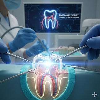
Tooth Pain Doesn't Have to Mean Tooth Loss
Root Canal Treatment Can Save Your Teeth
If you have severe tooth pain, sensitivity, swelling, or pain while chewing, a root canal may be able to save your natural tooth. At Vaishnavi Dental Clinic, we've helped patients preserve their teeth comfortably for over 24 years.
Root Canal Treatment is a specialized dental procedure designed to save a tooth that is severely decayed or infected.
When the inner part of your tooth—known as the pulp (containing nerves and blood vessels)—becomes inflamed or infected due to deep cavities or injury, a Root Canal is necessary to remove the damaged tissue and seal the space.
Are you experiencing these symptoms?

Severe Toothache
Is your tooth keeping you awake at night?
A persistent throbbing toothache, especially one that becomes worse at night, often suggests that the nerve inside the tooth is inflamed or infected. This type of pain rarely resolves on its own and usually requires professional evaluation.

Pain While Chewing
Does it hurt every time you bite down?
Pain while chewing may indicate a deep cavity, a cracked tooth, or an infection affecting the nerve inside the tooth. Ignoring it can allow the problem to worsen and make treatment more complex. A timely dental examination can help identify the cause and preserve your natural tooth whenever possible.

Gum Boil or Pimple Near Tooth
Do you have a small pimple or boil on your gums?
A recurring gum boil often indicates that infection is draining from the tip of the tooth's root. Even if the pain comes and goes, the underlying infection usually remains and should be evaluated by a dentist.

Broken or Dark Tooth?
A damaged tooth can expose the sensitive inner layers to bacteria, increasing the risk of infection. Early treatment may improve the chances of saving the tooth and restoring its strength and appearance.
How a Simple Cavity Becomes a Dangerous Infection?
A single tooth, left untreated, can progress through a series of increasingly painful and dangerous stages. This sequence illustrates exactly how that deterioration occurs, using illustrative animation.
1. The Initial Breach: Decay and Bacteria. We begin with the anatomy of a healthy human molar, shown in cross-section. The white enamel surface protects the sensitive interior. However, in this first image, we see the beginning of the end: a small, dark spot of Decay (1) on the enamel. A single, simplified purple Bacterium (2) approaches the spot, ready to invade. This small cavity is the entry point for the entire sequence.

The Deep Invasion: Pulp Infection
Building on the previous scene, the decay has now intensified. The small dark spot is a deep, dark brown cavity that has destroyed the enamel and the yellowish dentin layer beneath it. Crucially, the purple cartoon bacteria (established in the previous image) have formed a column or 'army' and have tunnelled deep into the tooth.
In this image, the bacteria are shown actively breaching the central Pulp cavity (the pink area where the nerves and blood vessels live). This causes the pulp—which was healthy and pink only moments ago—to become severely inflamed, changing to a deeper, angry red. The entire tooth structure is now compromised by this Pulp Infection.

The External Response: Pain and Swelling
At this stage, we pause the internal anatomy view (seen in Image 2) and shift perspective to visualize the symptoms. The pulp infection established previously has intensified and is now pushing outwards beyond the tooth roots.
We focus on the character's face. The character is visibly miserable: their eyebrow is tightly furrowed, and a tear is visible. Their left cheek is severely, exaggeratedly Swollen and rounded, a deep puffy red.
To connect this symptom back to the infection, we use a small circular inset. Inside this box, we visualize the Pain with a stylized, dark red pulsating abstract shape. Also visible in this inset are multiple copies of the identical purple Bacteria from the previous internal images, now clustered in a small pocket of yellow pus (an abscess) beneath the gum tissue. This connects the internal disaster to the external swelling.
The Final Disaster: Bone Infection
This is the peak of the bacterial invasion. We return to the internal anatomy cross-section (seen in Image 2), but pull the camera back significantly to show the entire tooth structure embedded within the surrounding jawbone (alveolar bone).
The disaster depicted in Image 2 (the breached pulp and bacterial column) is still active, but the infection has now migrated beyond the tooth entirely.
The army of purple Bacteria (which were seen clustering in the 'Pus Pocket' inset in Image 3) have now completely surrounded the root tips and are aggressively invading the surrounding bone tissue. The healthy, pink bone from Image 1 is gone; the jawbone here is mottled grey and sickly green, appearing 'moth-eaten' and deteriorated.
A large, oppressive, swirling vortex of purple bacteria and yellow-green pus occupies the deep bone space. The entire scene is bathed in a sickly, chaotic green light. This is Bone Infection (osteomyelitis).
.png)

Pain-Free Root Canal Treatment (RCT) in Vidyaranyapura, Bangalore
Your Expert Endodontic Care
Hello, I’m Dr. Sunitha Prasad, and with over 20 years of experience in dentistry, I've seen countless patients experience the discomfort of a compromised tooth. Often, a deep cavity, a crack, or even trauma can allow bacteria to reach the innermost part of your tooth, the dental pulp. This pulp contains nerves and blood vessels, and when it becomes infected or inflamed, it causes significant pain and, if ignored, can lead to serious long-term consequences.
Enhanced Precision
Rotary files, made of highly flexible nickel-titanium, navigate the natural curves of your root canals more accurately, ensuring thorough cleaning.
Reduced Treatment Time
The efficiency of rotary instruments significantly reduces treatment time, often allowing for single-visit root canal treatments in suitable cases.
Increased Patient Comfort: The smooth, continuous motion of rotary instruments creates less vibration and pressure compared to hand files, leading to a much pain-free RCT experience.
Predictable Outcomes: The consistent shaping and cleaning of root canals improve the chances of successful healing and long-term tooth preservation.
The Vaishnavi Dental Clinic Advantage:
Experience Meets High-Tech Care
My 20+ years of clinical experience, combined with state-of-the-art technology, ensures that your root canal treatment at our Vidyaranyapura clinic is of the highest standard.
We are committed to alleviating your pain and saving your natural tooth.
Expert Diagnosis & Planning
Every procedure begins with a thorough evaluation of your oral and general health. We utilize digital imaging to assess your bone, teeth, and associated structures, allowing me to plan the precise and safest approach for your RCT.
Advanced Equipment for Superior Results
Dentsply X-Smart Device: We leverage the Dentsply X-Smart Endodontic Motor, a high-end device that precisely controls the speed and torque of our rotary files. This ensures optimal cleaning, reduces procedural time, and significantly enhances patient comfort and precision during the entire process.
High-Quality Rotary Files: Our use of premium, flexible rotary files allows for meticulous shaping of even the most complex root canal anatomies.
Digital X-Rays:
Instant, low-radiation digital X-rays provide immediate diagnostic clarity.
Uncompromising Safety:
Your health is our priority. We adhere to a standardized 5-step sterilization protocol utilizing advanced autoclave sterilization and maintain rigorous hygiene standards throughout the clinic.
Comfort-Centric Ambiance:
From our friendly staff to our comfortable waiting areas, we strive to create a relaxing environment designed to ease any dental anxiety.
Seamless Logistics:
We maintain digital medical records for efficient access and follow-up, and offer multiple payment and EMI options to make quality care accessible.
The Root Canal Procedure: What to Expect
Thorough Evaluation: Initial consultation, digital X-rays, and often a CBCT scan in Vidyaranyapura to accurately assess the infection and anatomy.
Anesthesia:
Local anesthesia is administered to ensure a completely pain-free root canal treatment.
Access & Cleaning:
A small opening is made in the crown of the tooth to access the infected pulp. The Dentsply X-Smart device with specialized rotary files is then used to meticulously clean and shape the root canals.
Filling: The cleaned canals are disinfected and filled with a biocompatible material (gutta-percha) to prevent re-infection.
Restoration:
After the root canal, the tooth needs protection. We generally recommend an all-ceramic or Zirconium crown in Vidyaranyapura to reinforce the treated tooth, prevent fracture, and restore its natural appearance.
Post-Procedure Care & Long-Term Success
After your root canal treatment, a short healing period is expected. We provide clear instructions for post-operative care and schedule dedicated post-procedure follow-ups to monitor your progress and ensure the long-term success of your treated tooth. With proper care and regular check-ups at Vaishnavi Dental Clinic, your root canal treated tooth can last a lifetime!
Ready to Get Relief? Schedule Your Pain-Free RCT Consultation Today!
Don't let dental pain dictate your life. As the best root canal specialist in Vidyaranyapura, Bangalore, I am committed to providing you with gentle, efficient, and lasting solutions.